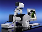
Definite Focus From Carl Zeiss Facilitates Research Of Cell Cultures Using A Microscope

With Definite Focus, Carl Zeiss offers a system for the Axio Observer.Z1 that can be used not only for the research of cell cultures in many biomedical applications, e.g. cell biology, pharmacology, molecular genetics, developmental biology and the neurosciences, but also in basic research.
The system has been specially designed for fluorescence applications and time-lapse experiments under live cell conditions. Definite Focus effectively counteracts drifting in the Z-direction. The microscopic image always remains in focus. Furthermore, preparation and waiting times in time-lapse experiments are markedly reduced. Since the introduction of the Axiovert 200 in 2000 and its successor, the Axio Observer, in 2006, hundreds of customers have successfully used these systems for long term time lapse imaging over periods as long as 72 hours. But increasing interest of integration of temperature impact on cell viability as part of the experiment put new demands on the ability of the imaging system to quickly compensate for temperature changes. Definite Focus helps solve this with its full integration into the workflow of the Axio Vision imaging workflow.
Definite Focus is coupled directly to the nosepiece carrier and directs infrared light of wavelength 835 nm into the beam path of the Axio Observer.Z1 inverted research microscope. To measure the distance between the objective and the specimen, a grid is projected onto the bottom of the culture vessel, and the reflection of this projection is analyzed. Possible deviations from the nominal value are determined and corrected quickly and very precisely via the Z-drive of the Axio Observer in the case of drifting. Even with high-magnification objectives, the system can be used without limitations for all contrasting techniques.
Definite Focus offers increased experimental reliability. Experiments that can no longer be evaluated due to drifting in the Z-direction are a thing of the past. Furthermore, incubation conditions can now be better structured according to needs. With stage incubation using the PM S1 incubator, for example, the user can quickly change the temperature as required during the experiment. Typical examples of dynamic temperature experiments include the analysis of protein folding mutants or heatshock experiments. Until now, performance of the relevant experiments had not been possible due to pronounced focus fluctuations.
Definite Focus can be operated directly via the TFT touchscreen display of Axio Observer.Z1. Convenient additional functions such as the storage of two nominal values and the possibility of using the system as a "focus finder" after replacement of the culture vessel are integrated into the system. Furthermore, Definite Focus is fully compatible with the AxioVision and LSM software from Carl Zeiss. The module supports all Axio Observer options such as TIRF, LSM or multiphoton microscopy. It works with all standard culture vessels, including multiwell dishes with glass or plastic bottoms. The supplied hardware comprises a sensor module on the nosepiece carrier, a beam combiner below the nosepiece and a Definite Focus controller.
About Carl Zeiss
Carl Zeiss MicroImaging, Inc., a subsidiary of Carl Zeiss, Inc., offers microscopy solutions and systems for research, laboratories, routine and industrial applications. In addition, Carl Zeiss MicroImaging markets microscopy and digital pathology systems for the clinical market, as well as spectral sensors for industrial and pharmaceutical applications. Since1846, Carl Zeiss has remained committed to enabling science and technology to go beyond what man can see. Today, Carl Zeiss is a global leader in the optical and opto-electronic industries.
With 11,249 current employees in the Group and offices in over 30 countries, Carl Zeiss is represented in more than 100 countries with production centers in Europe, North America, Central America and Asia. For more information on the breadth of solutions offered by Carl Zeiss MicroImaging, please visit www.zeiss.com/micro.
SOURCE: Carl Zeiss
